‘A perfect storm’
Cross-hospital collaboration helps a young French bulldog overcome a barrage of severe gastrointestinal problems that nearly turned deadly
Cross-hospital collaboration helps a young French bulldog overcome a barrage of severe gastrointestinal problems that nearly turned deadly
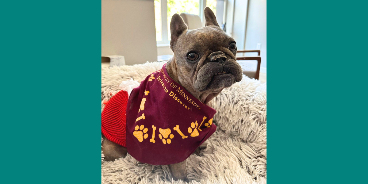
Hattie sits at home after a 15-day intensive care unit stay at the University of Minnesota Veterinary Medical Center.
Scroll through the Instagram account of 5-year-old French bulldog Hattie, and the photos paint a picture of a happy, snuggly, and feisty dog.
“She’s a little fireball who definitely has a mind of her own,” says Melissa Niebes, who’s had Hattie since she was a puppy.
But here and there, Niebes gives followers a glimpse into Hattie’s reality of living with multiple gastrointestinal conditions—a combination that almost took a deadly turn just over a year ago.
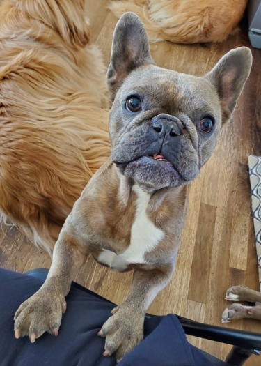
In a video clip, viewers can watch as Hattie eats a meal using a special device called a Bailey chair, which keeps her upright as she feeds. That’s because Hattie’s gastrointestinal problems impact her ability to move food down her esophagus, through her stomach, and to her intestines.
It took collaboration from experts from across the Emergency and Critical Care, Internal Medicine, Nutrition, and Surgery specialties at the University of Minnesota (UMN) Veterinary Medical Center (VMC) to diagnose, treat, and manage Hattie’s complex medical needs.
The trouble started the first week Hattie arrived home with Niebes. She refused to eat and landed herself in the emergency room overnight. Looking back, Niebes says the signs were there, but the full picture didn’t come together until Hattie’s symptoms began to escalate.
“As she continued to grow up, she was always really picky about food and not a food-motivated dog, which is unusual,” Niebes says. “A lot of times you had to trick her into eating and there was always a bit of stress around that.”
Prolonged bouts of vomiting and diarrhea coupled with weight loss brought Niebes and Hattie to the VMC’s Lewis Small Animal Hospital in Summer 2022. After exams and tests, Hattie was diagnosed with gastric reflux and esophageal dysmotility from continued acid reflux and vomiting, which means her esophagus is unable to exert proper muscle contractions to move food and water toward her stomach.
The Nutrition Service performed a motility study and prescribed alternative foods to help with dysmotility issues which appeared to be working as Hattie was getting better.
The situation took a hard turn and became dire when Hattie began having seizures due to an electrolyte imbalance. She spent 15 days in the hospital’s intensive care unit with clinicians across specialties working to keep her alive.
Upon examining Hattie, the Surgery Service identified thick scar tissue on the inside and outside of her pylorus—the lower portion of the stomach connected to the small intestine.
Hattie was diagnosed with pyloric stenosis, which means the muscles of her pylorus were abnormally thickened and prevented her stomach from emptying into the small intestine, causing food to back up into the esophagus.
The outside scar tissue was a congenital abnormality, and the internal scar tissue was caused over time by pushing food through the now inflexible pylorus. To improve Hattie’s health and quality of life, the surgeons recommended removing both sets of scar tissue. They performed the surgery and placed a gastrostomy (feeding) tube after Hattie struggled to eat.
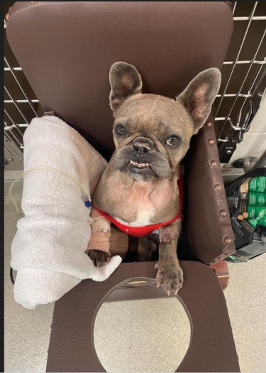
Dr. Zachary Lake, a resident in the Emergency and Critical Care Service, was part of the team helping treat Hattie during her extended stay in the ICU.
“In Hattie’s case, her main barrier was ensuring adequate caloric intake to allow her body to heal,” Lake says. “She was both not interested in taking in any food and would vomit or regurgitate anything that was supplemented to her.”
Due to the pyloric stenosis preventing food from entering the intestines, Hattie’s stomach had become dilated over time from being unable to empty itself.
“If something gets overly dilated for too long, then it doesn't work,” says Dr. Julie Churchill, head of the hospital’s Nutrition Service. “Her stomach was like a big saggy balloon. With this in mind, we had to try to figure out innovative ways to support her after her operation—it took some creativity among our teams.”
Because a traditional gastrostomy tube setup would not work with Hattie’s dilated stomach, the teams fashioned one that would bypass it and bring nutrition directly to her intestines.
Dr. Olivia Murray, who was a Small Animal Internal Medicine resident during Hattie’s treatment at the VMC, says gastrointestinal symptoms in dog breeds like French bulldogs aren’t uncommon, but that Hattie’s case was an example of a “perfect storm.”
This storm created a new reality for Niebes and Hattie after her discharge from the hospital.
Armed with a long list of feeding instructions, Niebes started their new routine of giving Hatti blended food through her tube.
“We were feeding her like five or six times a day in the very beginning,” Niebes says. “Between her medication schedule and her feeding schedule, it was like having a newborn. It was pretty intense for the first couple of months.”
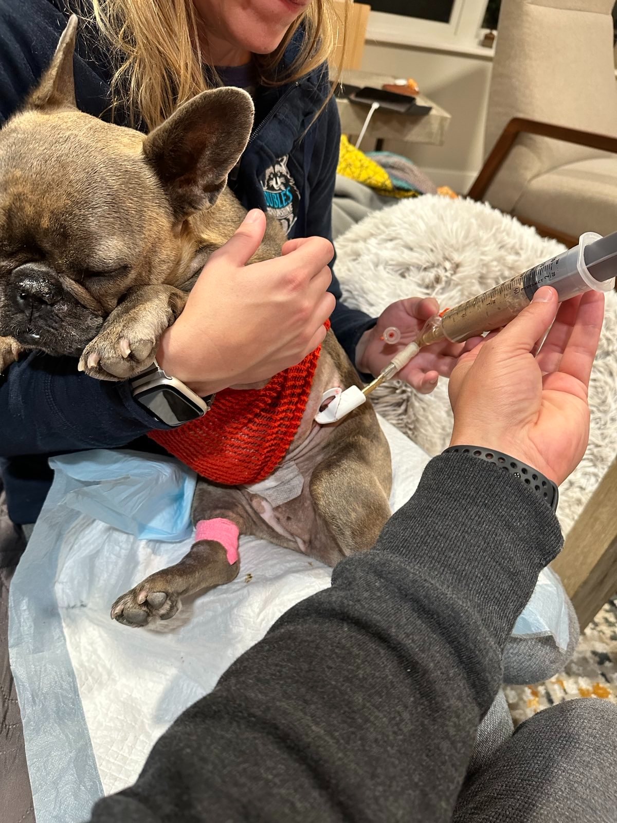
In time, Hattie’s stomach began to shrink back to a normal size, and she slowly began taking in food and sips of water through her mouth rather than her tube. During this time, Hattie needed to eat and drink while sitting up and remain so for 30 minutes after she finished her meal. Her frequent vomiting had damaged the sphincter between her esophagus and stomach. If she doesn’t stay upright, she risks regurgitating anything she ingests.
That’s where the Bailey chair comes into play. This special chair keeps Hattie’s esophagus in a position that allows food to move down it more effectively and prevents her from regurgitating.
After about six months after her ICU stay, Hattie’s feeding tube was removed. Murray credits the tube as an integral part of Hattie’s care and recovery.
“People are often averse to the idea of feeding tubes, however, they really can make recovery or management possible for the right cases,” she says. “Tube type, disease, and lifestyle factors all require careful consideration when recommending feeding tubes, but I would encourage owners to keep an open mind when the subject is broached.”
Hattie still eats meals in the Bailey chair, and Niebes works closely with the Nutrition team to continue managing Hattie’s diet. When she does come in for nutrition follow-ups or routine appointments, Hattie makes the rounds to visit the people who saved her life.
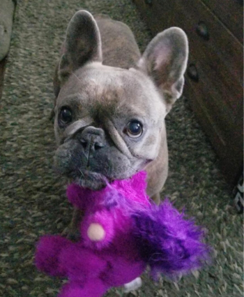
“Every time she comes in, we make sure that we take her to all those services, so they can see how great and healthy that is,” Churchill says. “That just really fills our cup and reminds us why we're here.”
The level of care Hattie received across the hospital’s services—especially during her ICU stay—is something Niebes says she is very grateful for. And despite the obstacles she’s faced, Hattie is living life to the fullest.
“We joke sometimes that she came back a different dog. She's so food-motivated now, which she just wasn't before because I'm sure she didn't feel well,” Niebes says. “She was eating to stay alive versus wanting to eat. That changed, and she's just more lively and happy now.”