Collaboration blasts glioblastoma
A five-year canine cancer research project makes for healthier dogs, happier owners, and—ultimately—healthier humans
A five-year canine cancer research project makes for healthier dogs, happier owners, and—ultimately—healthier humans
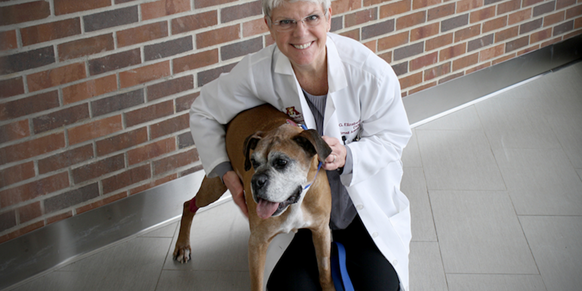
Photo by Amanda Stombaugh
The veterinary and human medicine communities have long observed that dogs and humans share a particularly deadly form of brain cancer known as glioblastoma. Not only do they have the disease in common, but humans and dogs also exhibit a similar immune response to its tumors. As G. Elizabeth Pluhar, DVM, PhD, DACVS, and her team work to manipulate this immune response and effectively kill glioblastomas in dogs, their work simultaneously carries over to advancing human medicine.
In its first year, Pluhar’s current project has already shown promise in improving the survival rates in dogs while also giving researchers a deeper understanding of glioblastoma as it applies to human trials. The $2.7 million grant—funded as part of the 21st Century Cures Act by the National Cancer Institute, part of the National Institutes of Health—is led by Pluhar, a professor of veterinary surgery in the Department of Veterinary Clinical Sciences at the University of Minnesota College of Veterinary Medicine (CVM).
“It’s exciting that this may be something that is immediately translated into human patients,” says Pluhar. “You know how we always talk about one year of human life equalling roughly seven years of canine life? Well, since our median survival time is now well over a year in the dogs we treat, that could translate into about five to seven years in people.”
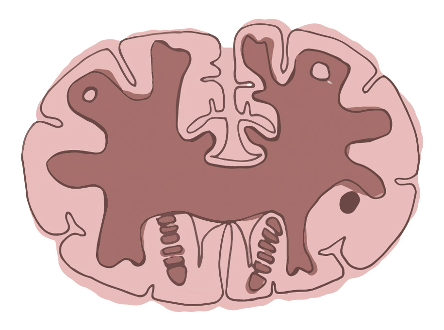
Glioblastomas are highly invasive tumors that carry a grim prognosis in humans, with a median survival of 14 to 16 months, despite intense treatment. Pet dogs diagnosed with these tumors have few options and are often euthanized shortly after diagnosis. Pluhar’s project, aiming to lengthen the life expectancy in dogs with glioblastomas, has combined complementary therapies in five dogs since funding for the trial began in 2017.
Now, Pluhar and her team are following the dogs that have had brain tumors surgically removed by performing magnetic resonance imaging (MRI) on each patient every four months to assess for tumor recurrence or progression. So far, the dogs that have had the first follow-up MRI’s have no evidence of tumor. With patients traveling to Minnesota from as far as Colorado and Canada to receive this innovative regimen, one thing is made clear: Pluhar’s work has made the U of M a destination for fighting this kind of cancer.
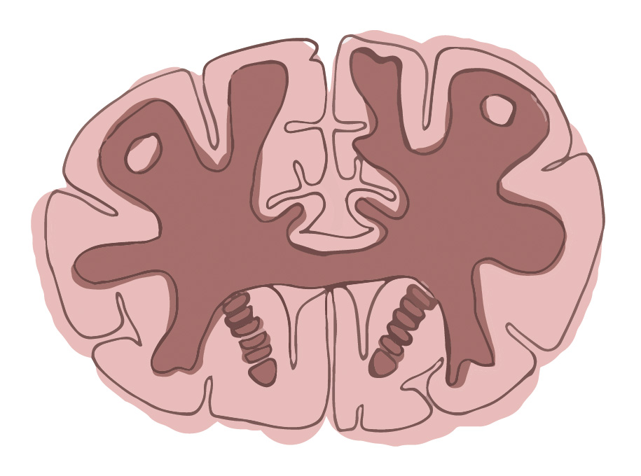
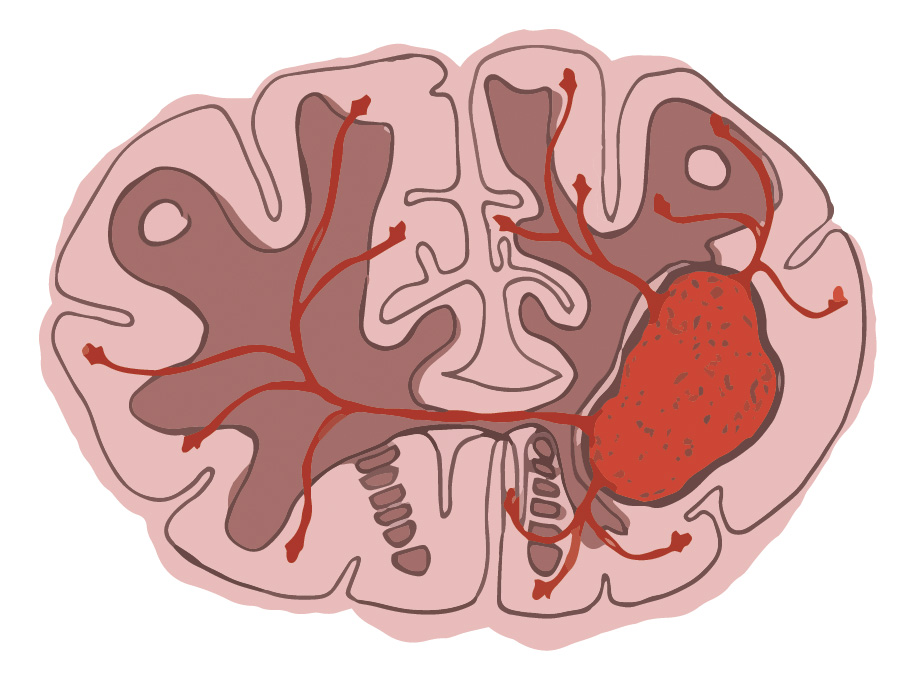
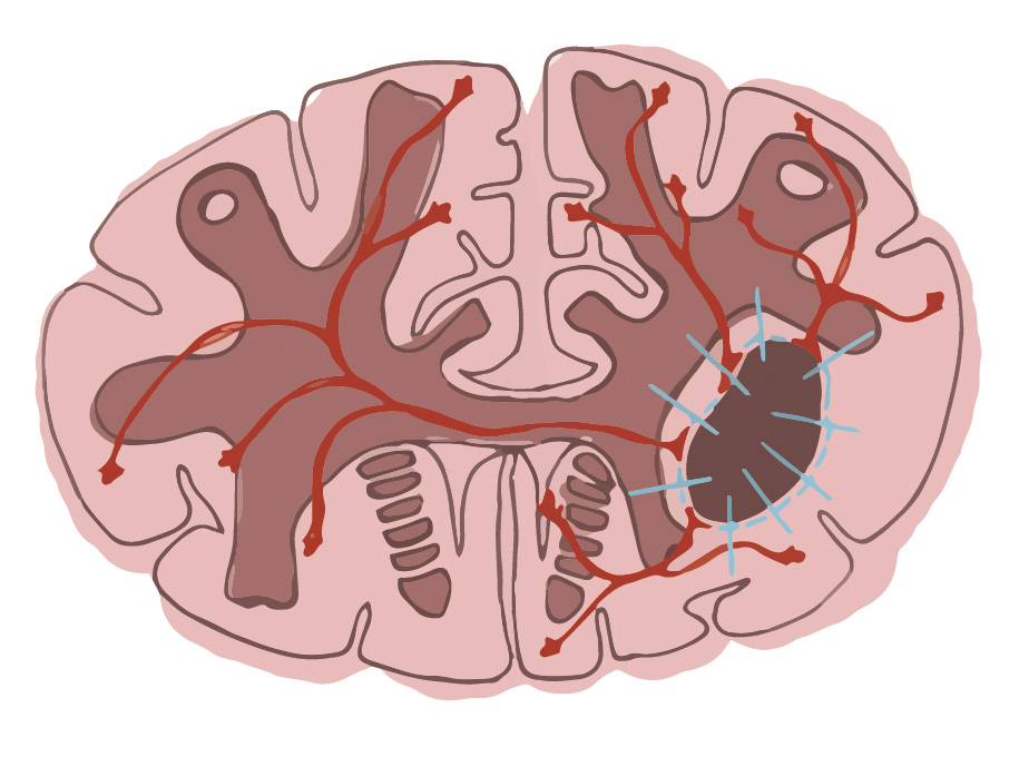
Illustrations by Megan Murrell
The new study represents a continued partnership between the CVM, the Masonic Cancer Center, University of Minnesota, the U’s Medical School, and the Medical School at the University of Michigan, to perform research on pet dogs with spontaneous tumors. With this collaboration has come additional support from the Randy Shaver Community and Cancer Research Fund, the American Brain Tumor Association, and the Humor to Fight the Tumor Foundation.
The group of researchers previously collaborated to use both vaccine-based and gene-based immunotherapy to treat dogs after surgical debulking of high-grade gliomas. These treatments had no adverse side effects and prolonged both progression-free and overall survival times more effectively than surgery alone.
Afterward, Christopher Moertel, MD, and Michael Olin, PhD, assistant professor in the Division of Pediatric Hematology and Oncology at the University of Minnesota, ran a small clinical trial in humans with recurrent glioblastoma, applying the same treatment.
“I have met some of the human patients that were treated in the vaccine clinical trial,” says Pluhar, “and they all said their quality of life was so good while undergoing immunotherapy treatment that they were able to do things they otherwise would not have been able to do had they continued on chemotherapy and radiation instead.”
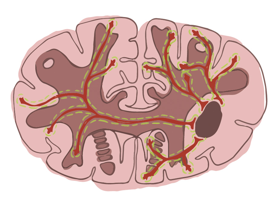
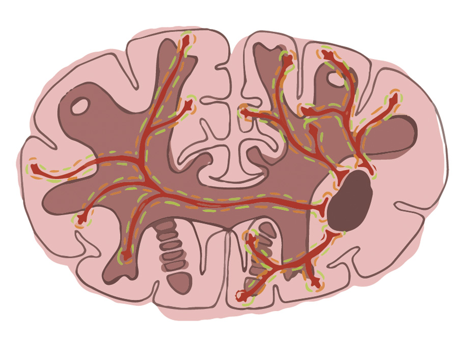
Although the previous approaches were successful at extending most of the canine patients’ survival, the tumors always recurred. To improve the efficacy of both therapies, Olin dove back in—he discovered that the tumors were elucidating a protein, CD200, that blocks the body’s natural immune response. So, Olin manufactured peptides of the native CD200 that override the tumor’s efforts to protect itself from the body’s immune response.
Olin found that mouse, canine, and human tumors all secrete this protein to thrive. As such, adding Olin’s CD200 peptides to immunotherapies brightens the future of treatment for both canines and humans alike. Olin’s contributions are not lost on Pluhar, who says the University’s collaborative culture is the backbone of the projects’ successes. “It’s just the atmosphere at the U of M, it’s expected that you are going to collaborate with people and it’s made easy to do here.”
Pluhar arrived at the University 15 years ago to work in orthopedics, which was her PhD’s focus. But since she had previous experience in neurosurgery, she and the late John Ohlfest, PhD, soon joined forces to shift cancer research from studying mice with lab-made tumors to dogs with spontaneously occurring tumors.
“We became one of the pioneers in canine neuro-oncologic surgery because John had this original idea to use dogs with spontaneous tumors as a model to assess therapies for people,” says Pluhar. “I thought at the time that we would just do an experiment in beagles and it would never go anywhere.” Pluhar has been pleasantly surprised to find that this work has actually professionally ensnared her.
And though she says retirement nears, Pluhar shows no signs of stopping. Her tank of fuel is full, in part due to the personal impact a close friend’s recent glioblastoma diagnosis has had on her. Watching her friend deal with the harsh aftermath of surgery, chemotherapy, and radiation propels her forward. “I know a lot of people with friends or family who are affected by these brain tumors,” she says. “It really goads me to continue this research because I think that our treatment approach is a really great alternative.”
The new research project hopes to demonstrate that combination immunotherapy, which will add the CD200 peptide with tumor lysate vaccines or local gene therapy, is safe and effective in pet dogs with spontaneous high-grade gliomas.
With so much promise on the horizon, Pluhar is quick to cite this project—and the ones that lead to it—as the highlight of her illustrious career: “Being involved in this has been very fulfilling—to know that what we have done has impacted both people and animals has just been amazing.”


