An ambidextrous approach
Better, cheaper canine cardiac care, right here
Better, cheaper canine cardiac care, right here
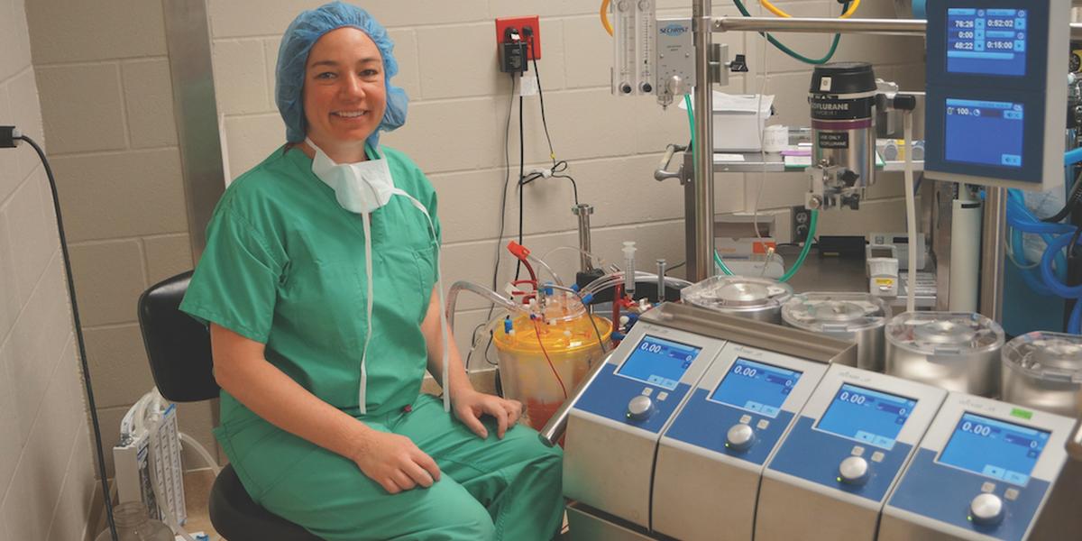
Photo courtesy of Wanda Gordon-Evans
Wanda Gordon-Evans, DVM, PhD, is performing groundbreaking surgery with one hand and finding ways to help owners save money with the other. Inspired to both borrow from and improve upon human medicine, Gordon-Evans was recruited from private practice to strengthen the University of Minnesota’s canine cardiac care team. Equipped with a research background in biomedical sciences, she aims to broaden the cardiovascular surgery program’s strong research and clinical foundations to benefit both pets and owners.
“I have always wanted to push veterinary medicine forward,” she says. “I was super happy to come back to academia and be involved in something that has potential to make a big scientific leap.” And when it comes to her work—specifically on mitral valve repair and replacement—Gordon-Evans’ innovative and resourceful mind-set is making her and her team a springboard for one such leap.
Mitral valve disease is an inevitability for many dogs over nine years old, and it typically results in a heart murmur that can be managed with medication. But for some less lucky canines, it can lead to heart failure. It runs most rampant in Cavalier King Charles spaniels, dachshunds, and Japanese Chins.
Though surgeons who perform canine mitral valve repair or replacement are rare, they usually use a similar method: clamping the patient’s aortic valve and submerging the patient’s heart in a solution that keeps it from beating. This method—called cross-clamping—has long been considered the gold standard in human medicine, providing the surgeon with a still, bloodless heart on which to operate. But returning a heart and its bodily home back to a stable state after this process is where complications and costs arise.
Cross-clamping often injures the heart because it results in a lack of blood circulating through the vital organ during surgery. The body can also become overly saturated with the fluids used to stop the heart, which means the patient will need a blood transfusion to bring their red blood cell count back to normal. Though these expensive post-op practices are commonplace for humans (and typically covered by insurance), they can ring up a rather high bill in a veterinary hospital. And for dogs, a hospital stay of as long as 12 days is needed to fully recover, which also adds to the bill for owners.
This entire medical endeavor typically adds up to around $20,000 for a dog owner—and that does not include the required travel to Japan, France, or the UK to have the surgery performed, since those are the only countries where it is routinely offered. As such, very few US dog owners are left with feasible options when their little Fido’s heart gives out from mitral valve deterioration.
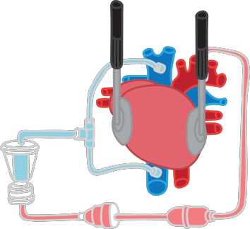
So, Gordon-Evans has retooled the process. Instead of cross-clamping, she uses electric shocks to strategically put her patient’s heart into fibrillation—a state that allows her enough stillness to rebuild a damaged mitral valve. Meanwhile, her patient is hooked up to a bypass machine so all organs can continue to be supplied with oxygenated blood. With the heart in fibrillation, Gordon-Evans only has to navigate a minor amount of blood to perform a repair, but the heart is still being oxygenated enough to avoid injury.
“Theoretically, if you are perfusing the heart with blood the whole time, there is less damage,” says Gordon-Evans, “so we should have less care needed afterward and certainly a faster recovery.” She also suspects that the dog’s stability following the procedure would call for a much shorter (and thus, cheaper) hospital stay of around three days. According to Gordon-Evans, humans could potentially undergo the same procedure.
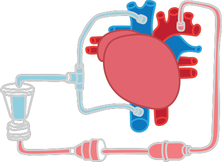
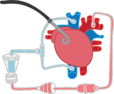
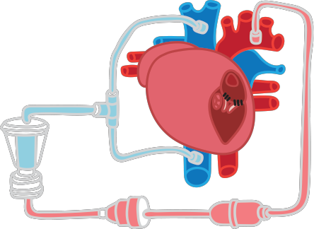
Illustrations by Megan Murrell
Gordon-Evans’ goal is multifaceted: not only is she thinking about the science, but she’s also laser-focused on improving the procedure’s price tag. She estimates her surgery would cost owners around $8,000, slashing more than half off the current price of a typical canine heart surgery.
Once Gordon-Evans has helped establish the U of M as the go-to place for canine mitral valve surgery in the United States, she has her sights set on expanding into congenital defects and even cats. “Humans and cats both suffer from hypertrophic cardiomyopathy,” she says. “Humans can have surgery and get off their meds.” Finding a way to apply this process to cats would be an opportunity to extend feline lives as well as to once again better human medicine’s approach.
Everyone—including dogs, cats, pet owners, and future human patients—will feel the reverberations of Gordon-Evans and her team’s work as their visionary advancement in cardiac surgery takes shape in the months and years to come.
But as always, Gordon-Evans remains steadfast in prioritizing affordability: “If we can make cardiac surgery more economical for owners, then it can be a turning point for heart disease.”
Making the procedure available to shelter animal patients. A fund previously established by the U of M’s Veterinary Medical Center—and supported by the generosity of donors—provides lifesaving, innovative care for homeless animals who otherwise might not receive these services and, upon recovery, make excellent candidates for forever homes. “For the very first cases we do,” says Gordon-Evans, “we would ideally provide the surgery at no cost to a humane society or rescue group.”
Starting with ownerless dogs has worked for Gordon-Evans in the past. She and her team recently treated a rescue dog whose blood was not circulating to his lungs. “We connected the aorta to the pulmonary artery so that more blood goes to the lungs,” she says. “He is doing fabulously; he just got adopted.”


