Ahead of the curve
Donor support elevates diagnostic imaging technology at the Leatherdale Equine Center to match the level of care its equine experts deliver
Donor support elevates diagnostic imaging technology at the Leatherdale Equine Center to match the level of care its equine experts deliver
The University of Minnesota College of Veterinary Medicine’s (CVM) Leatherdale Equine Center (LEC) now offers an equine standing computed tomography (CT) and neonatal intensive care unit (NICU). These new additions, made possible by a generous gift from Louise Leatherdale, elevate the level of care offered by LEC in multiple ways.

The new NICU allows LEC veterinarians to better offer around-the-clock care to sick and injured newborn foals. “There’s 11 months of caring for the mare and all of the expenses associated, so it’s heartbreaking when you see a foal fighting for its life,” Leatherdale says. The unit is set up with a large mare stall and a smaller, padded stall for the foal, along with state-of-the-art equipment. The two stalls are connected, with a sliding divider in place for use when the mare and foal need to be separated.
The pioneering standing CT was a major priority for Leatherdale, too. “The University and I have been in talks for years about how we can update, and we felt it was important to have the best diagnostics possible,” she says. “Horse owners in this community desperately needed it.”
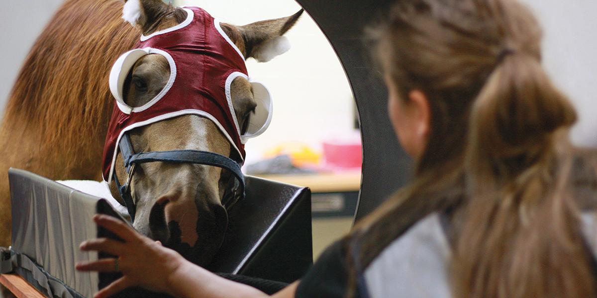
The standing CT, called "Equina" and developed by Asto CT, gives the LEC the region’s best suite of imaging capabilities. The CVM’s installation is only the second in the nation.
With the Equina, CVM veterinarians can quickly identify injuries to the head, neck, and legs without putting their patients through the stress of anesthesia. In addition to diagnosing head, neck, and limb injuries, the Equina can be used for full-body imaging of sedated foals and ponies to provide a 3-D perspective of areas such as the spine and pelvis.

“Having a standing CT allows us to more thoroughly image areas of the horse that we could only previously do with them under anesthesia,” says Troy Trumble, DVM, PhD, DACVS, associate professor in the Department of Veterinary Population Medicine (VPM). “For our clients, this technology will allow us to definitively diagnose lesions of the lower leg and head that we could not diagnose in the past, such as stress fractures.”
The new equipment better depicts the location and extent of an injury, which allows LEC veterinarians to develop better treatment strategies, prognoses, and surgical plans—especially for lower leg fractures or head and teeth issues, where the exact configuration or extent of injury is hard to define on traditional x-rays.
The standing CT played an imperative role in providing answers to Lori Zabel when her 11-year-old quarter horse, nicknamed Herb, was competing in a barrel racing event with Zabel and took a bad step around the third barrel. Herb held his right hind leg up in the air and refused to put any weight on it. Zabel’s veterinarian assessed Herb the next morning, and after nothing showed up on the x-rays of Herb’s hocks, she became concerned about a pelvic fracture.
Pelvic fractures can be life-threatening for horses, due to the pelvis’ proximity to main arteries. One of Zabel’s previous horses suffered a pelvic fracture that ended up severing an artery, so she knew Herb had to be examined with additional diagnostic equipment as soon as possible. Herb was transported within four hours to the LEC.
When we got to the U of M, the first person to greet us was Dr. Ernst, and it was his day off. That just speaks so highly of his dedication to the program and to his clients.
Lori Zabel
“When we got to the U of M, the first person to greet us was Dr. Ernst, and it was his day off,” says Zabel. “That just speaks so highly of his dedication to the program and to his clients.”
Upon arrival, Nicolas Ernst, DVM, MS, DACVS, associate professor in the Department of Veterinary Population Medicine, decided a bone scan was needed. Thankfully, the scan revealed an injury to the hock, not the pelvis. However, the scan didn’t reveal any specific fractures or soft tissue injuries, so the new standing CT was called into use.

“The CT was chosen because the radiographs were still unrewarding and there were no signs of soft tissue injury,” says Alyson Booth, DVM, a large animal intern at LEC. “The CT was able to show us what other diagnostics could not, which was the fracture lines on different planes.”
Luckily, the CT showed that the fracture in Herb’s hock was clean and wouldn’t require surgery. Herb is currently recovering at a rehabilitation facility in Nebraska. With a strict rehabilitation routine and three months of stall rest, Herb should make a full recovery and return back to barrel racing.
“It was amazing that the CT was able to show, in such detail, what the injury was,” Zabel says.
The LEC is distinctly situated to maximize the use of this new equipment to continue its tradition of pioneering world-class equine care. The equine center's specialist veterinarians have a longstanding history of innovating approaches to taking as close of a look as possible at equine patients, which they use to give a precise diagnosis and identify the most efficient and effective course of action.
Their expertise is augmented by many leading-edge technologies and tools, including a standing MRI that captures detailed images of the hoof, pastern, and fetlock; a dynamic overground endoscope that attaches to the horse’s bridle, is inserted into the nose, and feeds images wirelessly to a screen so veterinarians can assess respiratory issues in athletic horses as they move; and a gastroscope that allows for standing stomach examinations on even the largest patients.
The newest additions to this list of leading-edge tools, the standing CT and NICU are now available for patient use and are already adding a variety of health problems to the list of things within plain sight for veterinarians at the LEC. As the center broadens its technological reach to more effectively and efficiently find, tackle, and mitigate a myriad of equine health disorders, horses from around the country will continue to get back on their feet, faster.


